MBT Brackets Placements Technique PDF Free Download (Direct Link)
Traditionally, it has been recommended that pre-adjusted appliance brackets be placed with the twin bracket wings straddling, in a parallel fashion, the vertical long axis of the clinical crown, and that the center of the bracket slot be placed on the center of theclinicalcrown.1 Potential errors or potential deviations from his desired position can occurs follows: Horizontal bracket placement errors. These can normally be avoided with careful technique. Axial or paralleling bracket placement errors. These can normally be avoided with careful technique.
Excess bonding agent beneath the bracket base can cause thickness and rotational errors. Horizontal errors Brackets can be placed to the mesial or distal of the vertical long is of the clinical crown, leading to improper tooth rotation (fig. 1).Elimination of such errors can be best achieved by visualizing the vertical long axis of the crown directly from the facial surface, as well as from the incisal or occlusal surface with a mouth mirror. Some orthodontists even consider drawing a line through the vertical long axis of the clinical crown for more accurate visualization. Axial or paralleling errors Brackets can be rotated off the vertical long axis of the clinical crown if the bracket wings do not straddle the long axis of the crown in a parallel manner (fig. 2). Such errors lead to improper crown tip and can also be avoided by viewing the crown directly from the facial surface, as well as from the incisal or occlusal surface. Such errors can be eliminated by using the same techniques described for the elimination of horizontal errors.
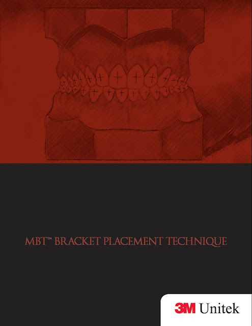
In This book the writer tells about the positions e.g horizontal, vertical and parallel brackets of teeth and their replacements techniques.
Direct visualization of the center of the clinical crown is a satisfactory method of locating this point on fully erupted and anatomically normal teeth. However in situations in
which there are gingival variations, differences in tooth size within the dentition, or incisal or occlusal variations, this direct visualization SUMMARY AND CONCLUSIONS
technique becomes more difficult.
Such situations do occur quite frequently in an orthodontic practice. A bracket placement chart was developed that allows the orthodontist to select a set of numbers representing the average center of the clinical crown for a given patient. Measurement gauges can then be used to check bracket positions after visual placement. The technique has been used in the practices of the authors for several months and has dramatically reduced the need for bracket repositioning due to incorrect visualization of the center of the clinical crown.
Thickness errors
Such errors can occur if excessive adhesive is left underneath one portion of the bracket base (fig. 3), or if the contour of the tooth does not correspond accurately to the contour of the base of the bracket.
Such errors can cause improper tooth torque or rotation, and can be eliminated by pressing the bracket against the tooth at placement, so that excessive adhesive flows from beneath the bracket, or by contouring the bracket base to more accurately fit the tooth surface.
Vertical errors
Vertical bracket placement errors occur when the bracket is placed gingival or incisal/occlusal to the center of the clinical crown (fig. 4).
Such errors lead to extrusion or intrusion of teeth, as well as potential torque and in/out errors.
The human eye is quite accurate at bisecting and locating the center
of a given object such as a crown, (as Andrews stated1). Therefore, brackets can be placed accurately using direct visualization on fully erupted and anatomically normal teeth. However, in the following clinical situations (which occur quite frequently),direct visualization is more difficult.
Book MBT Brackets Placements Technique is available to download free in pdf format.
- Name of Book: MBT Brackets Placements Technique
- Format: pdf
- Categories: Anatomy
- Publisher: 3M Unitek
- File Size: 3mb
Mccracken’s Removable Partial Prosthodontics 13th Edition PDF Free Download
MBT Brackets Placements Technique PDF Free Download
Alright, now in this part of the article, you will be able to access the free download of MBT Brackets Placements Technique using our direct links mentioned at the end of this article. We have uploaded a genuine PDF ebook copy of this book to our online file repository so that you can enjoy a blazing-fast and safe downloading experience.











![An Outline of Oral Surgery Part 2 by H.C. Killey (A Dental Practitioner Handbook) PDF Free Download [Direct Link]](https://www.cmecde.com/wp-content/uploads/2024/01/Download-An-Outline-of-Oral-Surgery-Part-2-by-H.C.-Killey-A-Dental-Practitioner-Handbook-PDF-Free-Direct-Link.jpg)
![Treatment Options Before and After Edentulism: Tooth Supported Overdentures 2023 Edition PDF Free Download [Google Drive]](https://www.cmecde.com/wp-content/uploads/2024/01/Treatment-Options-Before-and-After-Edentulism-Tooth-Supported-Overdentures-2023-Edition-PDF-Free.jpg)


