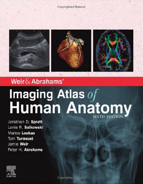
Weir and Abrahams’ Imaging Atlas of Human Anatomy PDF Free Download (Direct Link)
Imaging is ever more integral to anatomy education and throughout modern medicine. Building on the success of previous editions, this fully revised fifth edition provides a superb foundation for understanding applied human anatomy, offering a complete view of the structures and relationships within the body using the very latest imaging techniques.
It is ideally suited to the needs of medical students, as well as radiologists, radiographers and surgeons in training. It will also prove invaluable to the range of other students and professionals who require a clear, accurate, view of anatomy in current practice.
- Fully revised legends and labels and over 80% new images – featuring the latest imaging techniques and modalities as seen in clinical practice
- Covers the full variety of relevant modern imaging – including cross-sectional views in CT and MRI, angiography, ultrasound, fetal anatomy, plain film anatomy, nuclear medicine imaging and more – with better resolution to ensure the clearest anatomical views
- Unique new summaries of the most common, clinically important anatomical variants for each body region – reflects the fact that around 20% of human bodies have at least one clinically significant variant
- New orientation drawings – to help you understand the different views and the 3D anatomy of 2D images, as well as the conventions between cross-sectional modalities
- Now a more compete learning package than ever before, with superb new BONUS electronic enhancements embedded within the accompanying eBook, including:
- Labelled image ‘stacks’ – that allow you to review cross-sectional imaging as if using an imaging workstation
- Labelled image ‘slide-lines’ – showing features in a full range of body radiographs to enhance understanding of anatomy in this essential modality
- Self-test image ‘slideshows’ with multi-tier labelling – to aid learning and cater for beginner to more advanced experience levels
- Labelled ultrasound videos – bring images to life, reflecting this increasingly clinically practiced technique
- Questions and answers accompany each chapter – to test your understanding and aid exam preparation
- 34 pathology tutorials – based around nine key concepts and illustrated with hundreds of additional pathology images, to further develop your memory of anatomical structures and lead you through the essential relationships between normal and abnormal anatomy
Editorial Reviews
Review
About the Author
- Name of Book: Weir and Abrahams’ Imaging Atlas of Human Anatomy
- Format: pdf
- Categories: Anatomy
- Writer(s): Jamie Weir, Peter H. Abrahams
- File Size: 24mb
Viva in Dental Materials PDF Free Download
Weir and Abrahams’ Imaging Atlas of Human Anatomy PDF Free Download
Alright, now in this part of the article, you will be able to access the free download of Weir and Abrahams’ Imaging Atlas of Human Anatomy using our direct links mentioned at the end of this article. We have uploaded a genuine PDF ebook copy of this book to our online file repository so that you can enjoy a blazing-fast and safe downloading experience.

![An Outline of Oral Surgery Part 2 by H.C. Killey (A Dental Practitioner Handbook) PDF Free Download [Direct Link]](https://www.cmecde.com/wp-content/uploads/2024/01/Download-An-Outline-of-Oral-Surgery-Part-2-by-H.C.-Killey-A-Dental-Practitioner-Handbook-PDF-Free-Direct-Link.jpg)
![Treatment Options Before and After Edentulism: Tooth Supported Overdentures 2023 Edition PDF Free Download [Google Drive]](https://www.cmecde.com/wp-content/uploads/2024/01/Treatment-Options-Before-and-After-Edentulism-Tooth-Supported-Overdentures-2023-Edition-PDF-Free.jpg)


